
Demystifying Dental X-Rays: Are They Safe?
SMILE SMART READS
11/11/20245 min read
Understanding Dental X-Rays
Dental X-rays are a crucial diagnostic tool utilized in modern dentistry, enabling dentists to visualize the internal structure of teeth, roots, and surrounding bone. These imaging techniques are essential for identifying a variety of dental conditions that may not be readily apparent during a regular clinical examination. There are several types of dental X-rays, each serving distinct purposes in patient care.
Bitewing X-rays are one of the most common types, primarily used to detect cavities between teeth and assess the bone level around the teeth. They provide a quick view of the upper and lower teeth in one area of the mouth. Periapical X-rays, on the other hand, focus on the entirety of a single tooth, capturing both the crown and the surrounding bone structure. This type is particularly instrumental in diagnosing issues related to the roots of the teeth, including abscesses or other pathological conditions.
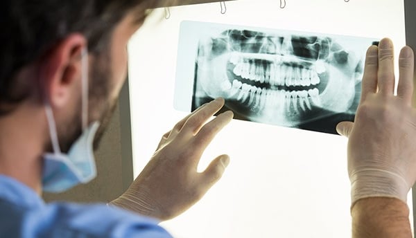
Another important type is the panoramic X-ray, which captures a wide view of the mouth in a single image, showing all teeth, jaw, and surrounding structures. This comprehensive view is indispensable for planning orthodontic treatment, detecting jaw disorders, or evaluating the condition of impacted teeth. The process of taking dental X-rays involves minimal radiation exposure, thanks to advancements in technology. Modern X-ray machines are designed to emit lower doses while still providing high-quality images for accurate diagnoses.
The benefits of dental X-rays extend beyond mere diagnostics; they also enhance treatment planning. For both patients and dentists, these imaging techniques are vital in recognizing issues that may require intervention, ensuring that timely and appropriate care can be administered. The integration of dental X-rays into routine dental visits underscores their importance in maintaining oral health and preventing complications.
The Science Behind Safety
The safety of dental x-rays has been a topic of discussion and concern among patients and healthcare professionals alike. To understand the safety aspects, it is important to consider the amount of radiation exposure involved in dental x-ray procedures. Generally, the radiation dose from a single dental x-ray is relatively low. For instance, a typical dental x-ray exposes a patient to approximately 0.005 mSv (millisieverts), which is comparable to the amount of radiation an individual absorbs from natural background radiation in about one day.
In comparison to other common sources of radiation exposure in daily life, dental x-rays are minimal. For example, a round-trip flight from New York to Los Angeles results in about 0.2 mSv of radiation exposure. Additionally, if one were to undergo medical imaging, such as a chest x-ray, the exposure might range from 0.1 to 0.2 mSv. Therefore, patients can feel reassured that the radiation dose associated with dental x-rays is significantly less than other activities and procedures they are likely to encounter.
Advancements in dental imaging technology have dramatically improved the safety profile of x-ray procedures. The shift from traditional film x-rays to digital x-rays, for instance, has reduced radiation exposure by up to 80%. Digital x-ray systems require a lower dose of radiation because they are more sensitive and produce clearer images more quickly. Furthermore, the introduction of lead aprons and thyroid collars as a protective measure has helped minimize exposure to surrounding tissues.
Health organizations, including the American Dental Association (ADA) and the Food and Drug Administration (FDA), have established rigorous guidelines governing dental x-ray practices. These protocols include recommendations for limiting the frequency of x-rays and implementing protective measures to assure patient safety. Overall, the evidence suggests that with proper protocols in place, the use of dental x-rays is safe and effective for oral health assessments and diagnosis.
Identifying Risks and Considerations
Dental X-rays are widely used diagnostic tools, enabling dentists to assess oral health and detect conditions that may not be visible during a standard examination. However, it is essential to consider the potential risks associated with their use, particularly for vulnerable populations such as pregnant women and young children. These groups require specific precautions due to the sensitivity of their developing bodies to radiation exposure.
For pregnant women, the potential risks posed by dental X-rays may involve concerns over radiation exposure to the developing fetus. To mitigate these risks, dental professionals typically recommend deferring non-emergency X-rays until after childbirth. If X-rays are necessary during pregnancy, special protective measures, such as the use of lead aprons and thyroid collars, are employed to shield the abdomen and thyroid gland from unnecessary radiation exposure. This proactive approach emphasizes the importance of informed consent and continuous communication between the patient and the dental team.
Young children are also at an increased risk when it comes to dental X-ray exposure due to their smaller size and developing tissues. Dentists assess the need for dental X-rays based on an individual child's dental history, growth patterns, and any specific dental concerns. Parents should engage in discussions with their dental care providers to ensure a clear understanding of the necessity of X-rays and the intended benefits, such as the early detection of cavities or developmental issues.
In most cases, dentists are diligent in evaluating each patient’s unique circumstances before recommending dental X-rays. They consider factors such as the frequency of dental visits, previous oral health issues, and the patient's risk factors for dental diseases. This careful assessment ensures that the diagnostic benefits provided by dental X-rays outweigh any potential drawbacks associated with their use, promoting a safe and effective approach to oral healthcare.
When Are Dental X-Rays Necessary?
Dental X-rays serve as an essential tool in modern dentistry, providing critical insights into a patient's oral health. They are particularly necessary during routine dental check-ups when a dentist needs to evaluate the overall condition of the teeth and gums. These examinations help identify potential issues that may not be visible during a standard visual inspection. Regular patients, typically, may require dental X-rays every one to two years, depending on their oral health status and risk factors for dental diseases.
In cases where tooth decay is suspected, dental X-rays become crucial for diagnosing the issue accurately. Early detection of cavities or decay through X-rays can lead to more effective treatment, preventing the problem from escalating into more severe conditions that necessitate extensive intervention. Furthermore, X-rays are indispensable for monitoring the progress of certain treatments, such as root canal therapy or periodontal treatment. This ongoing assessment ensures that the treatment is effective and helps in making necessary adjustments as needed.
Additionally, dental X-rays are often required before orthodontic treatments. They provide detailed images of the teeth alignment and jaw structure, aiding orthodontists in formulating a tailored treatment plan. For patients with specific health concerns, such as a history of dental issues or individuals with complex oral conditions, the frequency of X-rays may vary. It might necessitate more regular imaging to closely monitor changes in dental health.
Patients have the right to inquire about their dental X-ray requirements, and they can request alternative assessment methods where applicable. Open communication with healthcare providers ensures that patients can make informed decisions regarding their oral health, balancing the necessity of X-rays with their individual health considerations.